
Back pain: do I really need an MRI scan?
When is an MRI useful, why isn’t it always necessary, and what do its findings really mean?
When back pain appears, it’s natural to want to know “What’s wrong?” Many people assume that magnetic resonance imaging (MRI) is the best way to get a clear answer.
However, for most episodes of low back pain, having an MRI at the very beginning is neither necessary nor associated with better recovery.
This does not mean that the pain is not real or that it should not be taken seriously. It simply means that an image of the spine cannot always explain why someone is experiencing pain.
For this reason, current clinical guidelines recommend starting with a thorough clinical assessment: listening to the patient’s history, understanding how the pain started and how it has evolved, performing a physical examination and, when appropriate, a neurological assessment, while looking for any warning signs that may require further investigation.
An MRI is most useful when there is a specific clinical question to answer and when its result is likely to change the management of the condition.

Why isn’t an MRI recommended for every episode of back pain?
Most episodes of low back pain are classified as non-specific low back pain.
The term may sound surprising. It does not mean that healthcare professionals do not understand the problem or that the pain cannot be explained. It means that, after a thorough assessment, it is not possible to identify a single structure—such as a disc, joint or muscle—as the definite source of the pain. This is extremely common.
Low back pain is a complex experience. The tissues and structures of the back may contribute to it, but they are not the only factors that determine how much it hurts, how long it lasts or how much it affects everyday life.
That is why, in recent low back pain without red flags, routinely performing an MRI generally does not improve pain, function or recovery. In this early stage, a good clinical assessment usually provides much more useful information.
What can a healthcare professional assess without an MRI?
Much more than many people realise.
During the consultation and physical examination, it is possible to assess how the pain started, which movements or activities influence it, whether symptoms extend into the leg, whether there are changes in strength or sensation, and how the condition is affecting sleep, work and everyday activities.
When appropriate, a neurological examination can also be performed to assess muscle strength, sensation and certain reflexes.
One of the most important goals of this initial assessment is to identify any features suggesting a less common condition that may require medical assessment or further investigations.
When the medical history and physical examination are reassuring, treatment can usually begin without first obtaining an MRI of the spine.
Not having an MRI does not mean “doing nothing”
This is a particularly important point.
Sometimes, when a doctor or healthcare professional advises that an MRI is not indicated, patients may feel that their problem is not being investigated thoroughly.
However, deciding not to perform a test can also be a well-founded clinical decision.
If the assessment does not reveal concerning findings and the MRI is unlikely to change the treatment plan, avoiding an unnecessary test may actually be the most appropriate option.
In these situations, recovery usually focuses on maintaining or gradually regaining normal activities, temporarily adapting activities that significantly worsen symptoms, and progressively increasing movement and exercise.
At certain stages, relative rest may be helpful: temporarily reducing activities that clearly aggravate symptoms while avoiding, whenever possible, prolonged immobility or complete bed rest. The goal is not to push through any level of pain, but to find an amount of activity that is well tolerated and gradually increase it as recovery progresses.
When is a lumbar MRI actually indicated?
The most important question should not be: “Will the MRI show something?” Because it probably will.
The more useful question is: “Will the result of this MRI change what we do next?”
An MRI may be indicated when symptoms or clinical findings raise suspicion of a specific condition, when there are significant or progressive neurological deficits, or when recovery is not progressing as expected and the result is likely to influence treatment decisions.
It may also be appropriate after an adequate period of conservative treatment if a more specific intervention—such as an injection procedure or surgery—is being considered.
In these situations, the MRI is no longer simply a picture of the spine; it becomes a tool that answers a specific clinical question.
🏃 In practice
An MRI is not inherently good or bad. Its usefulness depends on when it is performed, the patient’s symptoms and, above all, whether the information it provides is likely to influence clinical decision-making.
What if I have sciatica? Do I need an MRI?
Not necessarily.
Pain that travels into the buttock or leg can be severe and worrying. In some cases, it may be related to irritation or compression of a spinal nerve root, for example because of a herniated disc.
However, having sciatica does not automatically mean that an MRI is needed straight away.
When there are no red flags or significant or progressive muscle weakness, clinical guidelines generally recommend starting with conservative treatment and monitoring how the condition evolves.
Many people improve gradually over the following weeks. In addition, the way symptoms evolve provides valuable clinical information.
If the pain persists, recovery is not progressing as expected, new neurological symptoms appear, or a more specific intervention is being considered, it may then be appropriate to reassess whether an MRI is needed.
🏃 In practice
The indication for an MRI can change over time. Just because an MRI is not necessary today does not mean it will never be needed.
What are the red flags for back pain?
Although the vast majority of episodes of low back pain are not caused by a serious disease, certain situations require prompt medical assessment. These are commonly known as red flags.
One of the most important warning signs is the development of new bladder or bowel problems, difficulty urinating, numbness around the genitals, perineum or inner thighs (sometimes called saddle anaesthesia), especially if accompanied by weakness in the legs. These symptoms may suggest significant compression of the spinal nerves, known as cauda equina syndrome, which requires urgent medical attention.
Prompt medical assessment is also recommended if there is significant muscle weakness or weakness that is getting worse over time—for example, if it becomes increasingly difficult to lift the foot or walk normally.
Other situations that may require further assessment include fever or chills associated with marked deterioration in general health, risk factors for spinal infection, significant trauma or suspected fracture, particularly in people with osteoporosis or increased bone fragility.
A history of cancer associated with new, persistent or unusual back pain should also be discussed promptly with a healthcare professional.
Having a red flag does not automatically mean that a serious disease is present. This distinction is essential. Red flags are not diagnoses.
Some are relatively common and, when considered in isolation, have limited ability to predict serious disease. For this reason, a good clinical assessment is much more than simply checking a list of symptoms. Healthcare professionals consider the whole picture: medical history, age, pain characteristics, symptom progression, the presence of several warning signs together and the findings of the physical examination. This clinical reasoning is what helps determine when an MRI is unnecessary and when it may become an important—or even urgent—investigation.
Why doesn’t an MRI always explain the source of back pain?
Why do abnormalities appear in people without pain?
This is probably one of the most important—and often surprising—concepts for understanding MRI findings of the spine.
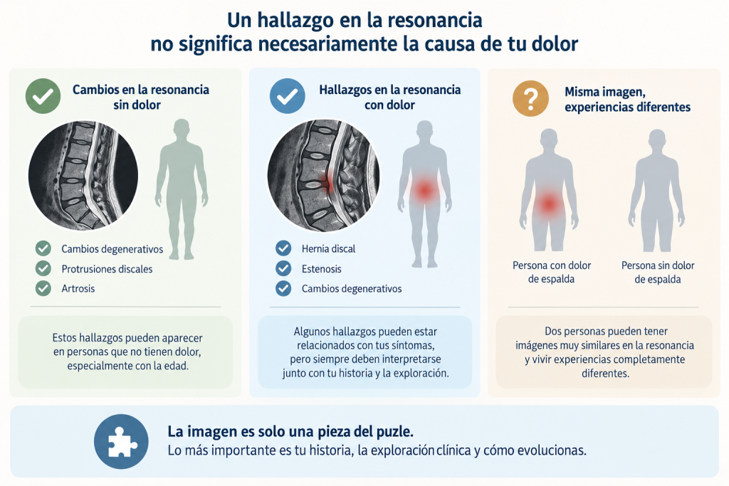
MRI is an extremely sensitive imaging technique. It provides detailed views of the intervertebral discs, facet joints, spinal canal, nerve roots and other spinal structures. And precisely because it reveals so much detail, anatomical changes are very commonly found.
The problem arises when these changes are automatically interpreted as injuries or assumed to be the cause of the pain.
Our bodies change as we age, and the spine is no exception. Intervertebral discs may gradually lose water content, change shape or develop small bulges. The facet joints may show signs of osteoarthritis, and other spinal tissues may also undergo progressive age-related changes.
Many of these changes are part of the normal ageing and adaptation process.
For this reason, what appears to be an “abnormal” finding on an MRI report can also be present in someone who has no back pain at all.
💡 Did you know?
A large systematic review analysing MRI and CT scans from 3,110 people without back pain found that degenerative changes were extremely common.
Disc degeneration was present in approximately 37% of people aged 20, increasing progressively to around 96% by the age of 80.
Disc bulges were also common: approximately 30% at age 20 and more than 80% by age 80.
Even disc protrusions could be found in young adults who had no symptoms at all.
These findings do not mean that MRI abnormalities are never important. They simply show that their presence alone does not prove they are the source of the pain.
A useful way to understand this is to think about wrinkles or grey hair.
They are visible signs associated with ageing, but we do not automatically interpret them as a disease.
The comparison is not perfect, but it illustrates a key principle: a structural change does not necessarily mean tissue damage, fragility or pain.
My MRI says “disc degeneration”, “disc protrusion” or “osteoarthritis”. Should I be worried?
Not necessarily.
Radiology reports use medical terminology to describe what is visible on the images. Terms such as degenerative disc disease, disc dehydration, disc protrusion, disc bulge, facet joint osteoarthritis and degenerative changes can sound alarming when read without context.
However, these terms primarily describe anatomical features rather than a diagnosis or the definite cause of pain.
They do not automatically indicate how much pain a person should experience, how well they should be able to move or how their condition will progress.
For example, degenerative disc disease describes changes within an intervertebral disc. A disc protrusion means that part of the disc extends beyond its usual boundaries. Osteoarthritis describes specific changes affecting a joint.
All of these findings may contribute to some people’s symptoms. But they can also be present without causing any symptoms at all.
That is why an MRI report should never be interpreted in isolation.
🏃 In practice
An MRI report should be interpreted, not simply read.
Its meaning depends on whether the findings are consistent with the location of the symptoms, the clinical examination, the neurological assessment and the patient’s overall course.
“My MRI shows a herniated disc. Does that mean it’s causing my pain?”
It might. But not always.
A herniated disc can irritate or compress a nerve root and produce characteristic symptoms, such as pain radiating down the leg, altered sensation and, in some cases, muscle weakness. When the location of the herniation matches both the symptoms and the neurological examination, MRI findings can provide very valuable information.
This is what we call clinical-radiological correlation.
However, herniated discs and disc protrusions can also be found in people who have no pain at all.
So it is not enough to ask, “Is there a herniated disc?”
We must also ask, “Does this herniated disc actually explain what this person is experiencing?”
This distinction may seem subtle, but it fundamentally changes how an MRI should be interpreted.
Does a herniated disc mean my spine is permanently damaged?
No. This is another common misconception that can create unnecessary concern.
A herniated disc is not necessarily a structure that will remain unchanged for the rest of your life. The body has the ability to adapt and remodel tissues over time.
In fact, numerous studies have documented the spontaneous regression or resorption of herniated discs during conservative treatment.
This may seem surprising: some herniated discs that are clearly visible on an initial MRI become much smaller—or may even no longer be visible—on later scans.
Spontaneous resorption appears to be particularly common in extruded and sequestered disc herniations.
This does not mean that every herniated disc disappears or that they all follow the same course.
But it does challenge an important misconception: a herniated disc should not automatically be interpreted as irreversible damage to the spine.
And there is another even more important point.
The evolution of MRI findings and the evolution of pain do not always go hand in hand.
A person may recover significantly even though certain MRI findings remain unchanged. Likewise, improvements on imaging do not necessarily lead to proportional improvements in symptoms.
Our clinical goal, therefore, is not necessarily to achieve a “perfect” MRI, but to help people regain function, movement, confidence and quality of life.
Imaging is not the same as pain
This idea captures a large part of the issue.
For many years, musculoskeletal pain was explained primarily through a structural model:
something is damaged → that damage causes pain → the damage must be corrected to eliminate the pain.
In some situations, this model works very well. A fracture, an infection or certain types of neurological compression have a clear structural basis.
But for many people with low back pain, reality is more complex.
There is a relationship between certain MRI findings and pain, but it is not absolute.
Some people have major structural changes with very few symptoms, while others experience severe pain despite having few visible abnormalities on imaging.
This does not mean the pain is imaginary.
It means that pain cannot be explained solely by what we see on an image.
Can having an MRI too early make things more complicated?
In certain situations, yes.
Not because MRI itself is dangerous—unlike X-rays or CT scans, MRI does not use ionising radiation—but because of the consequences of identifying findings that may not be clinically relevant.
Imagine someone develops a recent episode of low back pain without any red flags.
An MRI is performed, and the report describes multilevel degenerative disc disease, a disc protrusion and arthritic changes.
Even if these changes had been present for years before the pain began, the person may interpret the results as: “My back is damaged.”
From that point on, they may become afraid to bend, lift, exercise or even perform everyday movements.
In some cases, these findings may also lead to additional tests, more medical appointments or interventions that might never have been necessary.
This phenomenon is part of what we know as overdiagnosis and low-value healthcare.
💡 Common myth
“The more tests I have, the better we’ll know what’s wrong.”
Not always. A test is truly useful when it answers a specific clinical question and helps guide a better decision.
More information does not always mean more useful information.
Does this mean MRIs are a bad thing?
Not at all. The goal is not to move from ordering too many MRIs to distrusting them.
MRI is an outstanding diagnostic tool and can be essential in certain situations.
The problem is not the MRI itself.
The problem arises when an image is interpreted outside its clinical context or when a scan is requested without a clinical question that could change management.
When appropriately indicated, MRI can help identify specific conditions, assess neurological structures, plan procedures or complement a clinical assessment.
The key is to use it for the right patient at the right time.
Understanding back pain beyond MRI: the biopsychosocial model
If MRI alone cannot fully explain pain, a logical question follows: where does pain come from?
Current scientific evidence suggests that the answer is more complex than identifying a single structure.
Pain is a real experience generated by the nervous system from multiple sources of information.
Tissues certainly matter.
Nerve irritation, a recent injury, physical capacity, certain movements, inflammation or the sensitivity of specific tissues can all contribute to the pain experience.
But other factors also play a role.
Sleep, stress, previous pain experiences, worries, fear of movement, emotional state, physical activity, work demands and the social environment can all influence how we experience pain and how we recover.
This is what we call the biopsychosocial model of pain.
These factors can be grouped into three broad dimensions:
- Biological: tissues, muscles, joints, discs, the nervous system, physical fitness, sleep and general health.
- Psychological: worries, stress, fear of movement, expectations, previous experiences and coping strategies.
- Social: work, family, environment, daily activities, social support and personal circumstances.
These dimensions do not function independently. They constantly interact with one another.
This does not mean that every episode of pain has a psychological cause.
It means that we are people, not MRI scans.
💡 Common myth
“If stress influences my pain, it means the pain is all in my head.”
No. Pain is a real experience.
The fact that stress, sleep, emotions or context can influence pain does not make it any less real. In the same way, stress can affect heart rate, digestion or muscle tension without making any of these phenomena imaginary.
Understanding this biopsychosocial dimension broadens the range of treatment options.
Instead of trying to identify a single structure that needs to be “fixed”, we can identify which factors are modifiable for that specific person.
That leads us to a much more useful question than “What does my MRI show?”:
“What can I do to gradually regain my activities and rebuild confidence in my back?”
What should I do when an MRI is not recommended?
Not needing an MRI does not mean you have to wait passively for the pain to disappear.
For most episodes of low back pain without red flags, one of the main goals is to maintain—or gradually regain—movement and everyday activities.
This does not mean ignoring pain or forcing movements that are clearly intolerable.
During the first few days, it may be helpful to temporarily adapt certain activities. This is known as relative rest: reducing activities that clearly aggravate symptoms while maintaining, as much as possible, the movements and activities that are well tolerated.
Walking, changing position regularly and continuing with some daily activities are often preferable to remaining still for long periods.
As symptoms allow, the goal is to progressively increase activity.
For some people, clear explanations and staying active will be enough. For others, an individualised exercise programme, physiotherapy or other strategies adapted to the factors contributing to the pain may be beneficial.
When pain persists, it may also be important to explore factors such as sleep, stress, fear of movement, work demands or a loss of physical conditioning.
There is no single approach that works for everyone.
Should I wait until the pain has completely gone before moving again?
Generally, no.
One of the beliefs that most commonly limits recovery is thinking that pain always means damage and that any painful movement is causing further injury.
With some acute injuries, temporarily protecting a structure may indeed be necessary. However, for many cases of low back pain—especially once serious conditions have been ruled out—a certain degree of discomfort during recovery does not necessarily mean damage is occurring.
For this reason, returning to activity can usually be done progressively by temporarily adjusting the intensity, duration and frequency.
The goal is not to choose between two extremes—complete bed rest or ignoring the pain—but to find a level of activity that your body can tolerate and gradually build upon.
🏃 In practice
The question is not always: “Can I do this movement without feeling anything at all?”
Sometimes a more useful question is: “Can I do this movement at a tolerable level and recover well afterwards?”
What role do exercise and physiotherapy play?
Exercise can play an important role, especially when pain persists or limits daily activities.
However, there is no single “perfect exercise” for back pain.
Walking, strength training, mobility exercises, aerobic exercise or programmes that combine different types of activity may all be beneficial, depending on the individual.
The important thing is to find an activity that can be performed consistently and progressed according to the person’s individual response.
In this context, physiotherapy should not be limited to trying to “correct” a structure.
It can help improve mobility, strength and tolerance to specific activities, while also helping people better understand their symptoms and gradually progress towards meaningful goals: returning to running, lifting a child, working without fear, training in the gym or simply carrying out daily activities with greater confidence.
What about manual therapy?
Manual therapy can be part of the treatment strategy for some people, especially when it helps temporarily reduce symptoms or makes movement easier.
However, it should not reinforce the idea that the spine constantly needs to be “put back into place” or that the body depends on a healthcare professional to function properly.
Its role should be to facilitate an active recovery—not replace it.
In many cases, combining education, progressive movement, exercise and selected manual therapy techniques is more consistent with current evidence than relying on a single treatment in isolation.
How do we manage back pain at Aliantis?
At Aliantis, we try not to reduce the problem to a single question: “Which structure is damaged?”
Instead, we broaden the assessment by asking: “What could be contributing to this person’s pain, and what do they need to recover their activities?”
The first step is listening carefully to the person’s history and performing an appropriate clinical examination. We assess movement, strength and function and, when symptoms require it, we perform a neurological examination. We also consider how pain affects sleep, work, sport and everyday life.
A fundamental part of the assessment is determining whether there are any findings that make medical evaluation or additional investigations advisable.
When the situation is reassuring, the goal is to build a progressive recovery strategy.
It may include pain education, recommendations to stay active, exercise, physiotherapy and, when appropriate, manual therapy.
In more complex or persistent situations, our multidisciplinary approach also allows us to consider other factors that may be influencing recovery.
The ultimate goal is not for someone to need treatment indefinitely.
It is to provide people with the knowledge and tools to better understand their symptoms, gradually regain their abilities and become more confident and independent in managing their health.
So, do I really need an MRI if I have back pain?
For most recent episodes of low back pain without red flags, not immediately.
An MRI can be an extremely valuable tool when there is a specific clinical suspicion, important or progressive neurological deficits, or when the result is likely to change treatment decisions.
However, MRI scans can also show disc protrusions, disc degeneration, osteoarthritis and other changes in people who have no pain at all.
For this reason, imaging findings should never be interpreted in isolation.
An MRI shows anatomy. It does not directly measure how much pain someone feels, what movements they can perform or how they are likely to recover.
Clinical assessment is what brings all those pieces together.
And in many cases, when there are no red flags, the best starting point is not to immediately look for a “damaged” structure, but to understand the problem, maintain a tolerable level of activity and gradually rebuild movement, strength and confidence.
The best MRI is not the one performed “just in case”.
It is the one performed at the right time, to answer a specific clinical question and when the result can genuinely help guide better decisions.
Conclusion
Having back pain does not necessarily mean you have a serious injury, and finding an abnormality on an MRI does not automatically identify the cause of your pain.
Modern healthcare aims to avoid two extremes: performing imaging routinely for everyone, or assuming it is never necessary.
The key is to use imaging when it is likely to provide information that is genuinely useful.
When there are no red flags, a thorough clinical assessment, clear information, relative rest when appropriate and a gradual return to movement and daily activities are often an excellent starting point.
If, during recovery, new symptoms appear or the clinical situation changes, an MRI may then fulfil its real purpose: helping clinicians make a better-informed decision.
MRI helps us understand the spine. Clinical assessment, scientific evidence and dialogue help us understand the person. And that is where truly personalised care begins.
Frequently Asked Questions about MRI and Back Pain
When should I have an MRI for low back pain?
In most cases, an MRI is not needed immediately for recent low back pain without red flags.
It may be appropriate when there is suspicion of a specific underlying condition, important or progressive neurological deficits, or when the result is likely to change treatment decisions.
The decision should be based on the clinical history, examination findings and symptom progression—not simply on how intense the pain is.
Do I need an MRI if I have sciatica?
Not always.
Sciatica often improves with conservative treatment without the need for immediate imaging.
MRI becomes more relevant when there is significant or progressive muscle weakness, red flags, symptoms are not improving, or when an intervention is being considered.
What can a lumbar MRI show?
An MRI provides detailed images of the intervertebral discs, nerve roots, spinal canal, facet joints and other spinal structures.
It may reveal disc herniations or protrusions, degenerative changes, spinal stenosis and other structural findings.
However, many of these findings are also common in people without pain and should always be interpreted alongside symptoms and the clinical examination.
Is a disc protrusion or herniated disc on MRI serious?
Not necessarily.
Its significance depends on its location, the type of herniation and, most importantly, whether it matches the person’s symptoms and neurological examination.
A disc herniation may be clinically important in one person but simply an incidental finding in another.
Having a disc herniation does not automatically mean the spine is fragile or permanently damaged.
Can a herniated disc disappear?
Yes. Disc herniations can decrease in size or even regress spontaneously over time.
Scientific studies show that this is relatively common, particularly with extruded or sequestrated disc herniations.
Even when the MRI appearance does not completely normalise, many people recover very well with appropriate conservative management.
Which is better for back pain: an X-ray or an MRI?
It depends on what you are trying to assess.
An X-ray mainly allows healthcare professionals to assess bony structures and can be useful in certain situations, for example when a fracture is suspected.
MRI provides much more detailed information about discs, nerves and soft tissues.
However, for non-specific low back pain without red flags, the most important question is usually not which imaging test to choose, but whether any imaging is actually needed at all.
This blog article does not aim to generate new knowledge; it is based on the reading of scientific publications, blog articles and other texts.
Sources:
National Institute for Health and Care Excellence (NICE). Low back pain and sciatica in over 16s: assessment and management (NG59). Recommendations on assessment, imaging, self-management and treatment.
World Health Organization (WHO). WHO guideline for non-surgical management of chronic primary low back pain in adults in primary and community care settings. Geneva: WHO; 2023.
American College of Radiology (ACR). ACR Appropriateness Criteria®: Low Back Pain. Revised 2021.
Brinjikji W, Luetmer PH, Comstock B, et al. Systematic literature review of imaging features of spinal degeneration in asymptomatic populations. AJNR Am J Neuroradiol. 2015;36(4):811–816.
Chou R, Fu R, Carrino JA, Deyo RA. Imaging strategies for low-back pain: systematic review and meta-analysis. Lancet. 2009;373(9662):463–472.
Shraim BA, Shraim MA, Ibrahim AR, et al. The association between early MRI and length of disability in acute lower back pain: a systematic review and narrative synthesis. BMC Musculoskelet Disord. 2021;22:983.
Zou T, Liu XY, Wang PC, et al. Incidence of spontaneous resorption of lumbar disc herniation: a meta-analysis. Clin Spine Surg. 2024;37(6):256–269.
Rashed S, Vassiliou A, Starup-Hansen J, Tsang K. Systematic review and meta-analysis of predictive factors for spontaneous regression in lumbar disc herniation. J Neurosurg Spine. 2023;39(4):471–478.
World Health Organization. Low back pain – Fact sheet. WHO. Updated overview of low back pain, clinical assessment, rehabilitation and biopsychosocial management.